- Home
- left breast bigger than right breast
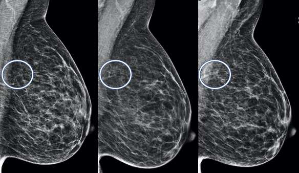
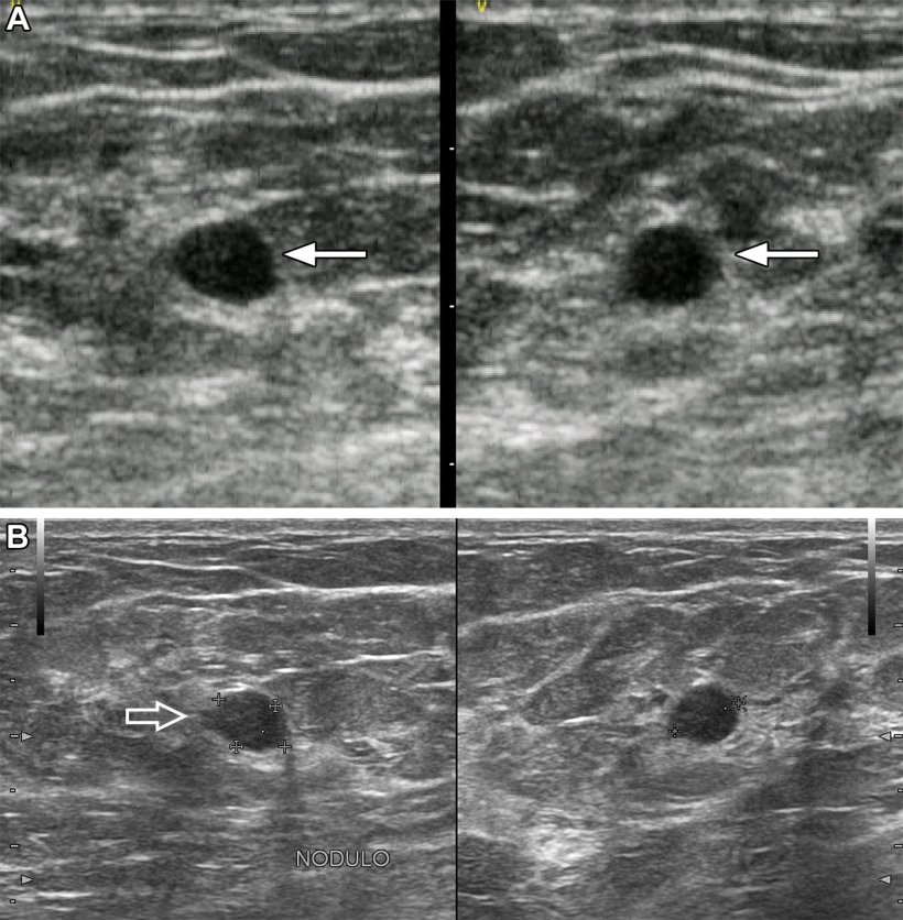
- A 60-year-old patient presented by a lump in the left breast
A 60-year-old patient presented by a lump in the left breast
4.8 (612) · $ 18.50 · In stock

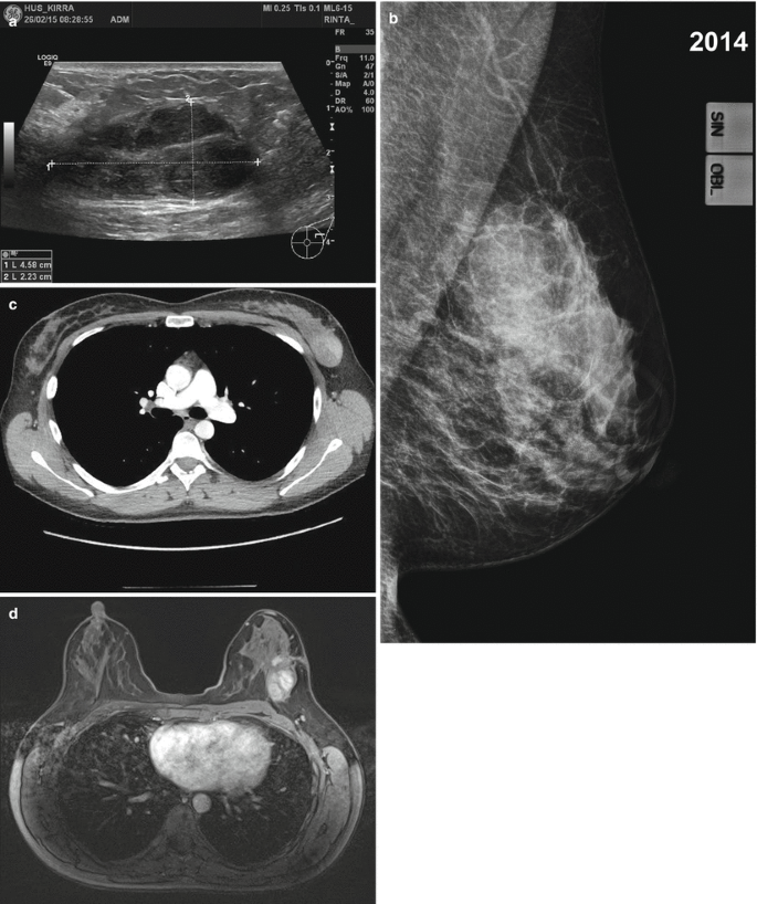
Download scientific diagram | A 60-year-old patient presented by a lump in the left breast. Mammography revealed focal asymmetry in the left upper inner quadrant with microcalcifications (a, b). DBT showed left breast spiculated mass with microcalcifications as well as right breast retroareolar nodule with microcalcifications (c, d). CEM showed left breast heterogeneously enhancing upper inner quadrant mass lesion with spiculated margins and surrounding multiple satellite lesions as well as right breast tiny right retroareolar homogenously enhancing mass with not circumscribed irregular margins (e, f). Breast ultrasound showed left breast irregular ill-defined mass in the left upper inner quadrant as well as right retroareolar small irregular ill-defined mass (g, h). The final diagnosis was bilateral invasive duct carcinoma from publication: Comparative study between contrast-enhanced mammography, tomosynthesis, and breast ultrasound as complementary techniques to mammography in dense breast parenchyma | Background Mammography is accused of having low sensitivity and specificity in dense breast parenchyma. Also, women with dense breasts show an increased risk of developing breast cancer. Breast ultrasound has been used for several years for a better characterization of breast | Breast Ultrasound, Mammography and breast | ResearchGate, the professional network for scientists.
AI helpful in triaging breast masses in low-resource areas •

A 60-year-old woman, presented with a palpable left breast mass; (a)

Breast Cancer Patient Develops Heart Palpitations and Dyspnea

/files/Articles/976947/fonc-12

Heba Azzam's research works Cairo University, Cairo (CU) and other places
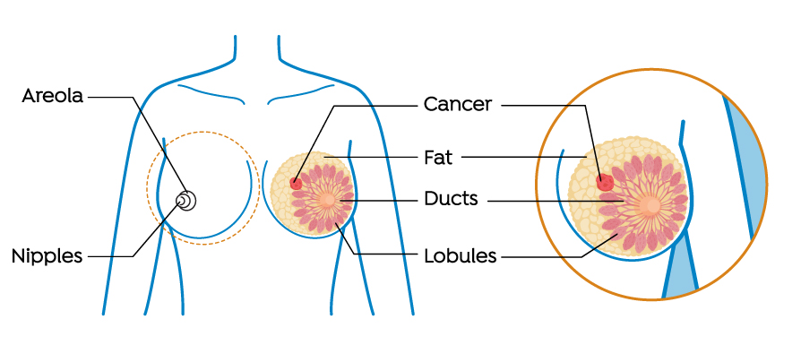
Patient education - Breast cancer - UF Health

Chest Wall Tumour: Causes, Symptoms, Signs, and Treatment - MyHealth

Breast cancer - Wikipedia
A Guide to Breast Cancer in Older Adults
Sarcoma of the Breast