Cell and flare in the eye (Video)
5 (458) · $ 7.00 · In stock
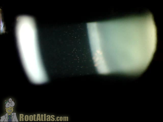
This video demonstrates what cell and flare look like under the slit-lamp microscope. “Cell” is the individual inflammatory cells while “flare” is the foggy appearance given by protein that has leaked from inflamed blood vessels. This finding is commonly seen with uveitis, iritis, and after surgery … and actually seeing it can be challenging for

Quiz: Persistent Corneal Edema – Why? – Cataract Coach™

Monday Back to Basics
Iris Beam's Instagram, Twitter & Facebook on IDCrawl

Sun

Cells and Flares, White blood cells and/or Proteinaceous fluid in the anterior chamber; known as “cell and flare” Aqueous Cells Anterior chamber cells are primarily

South Alabama Emergency Medicine (@SouthEmergency) / X

Videos

Tugas Modul Gangguan Mata Tutor 10

Flare sign in Ophthalmology. Grading of Ocular Inflammation.

Videos
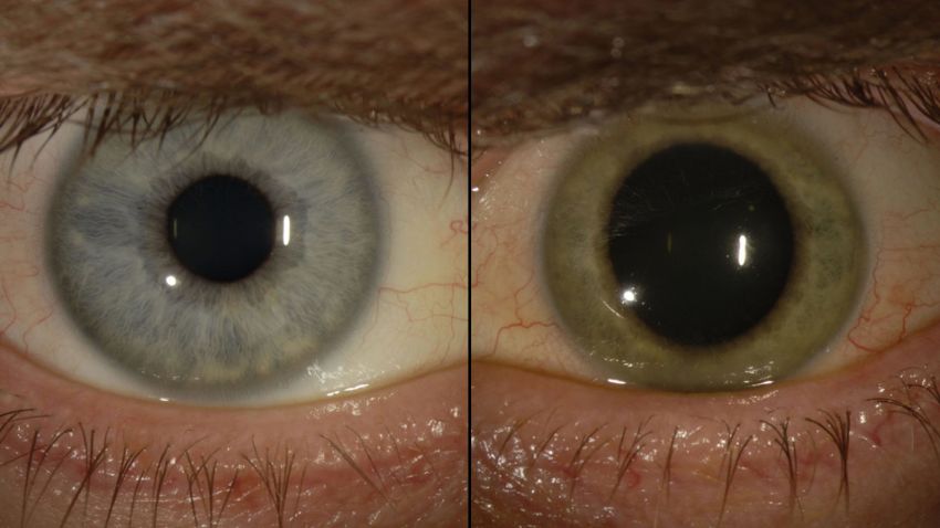
American doctor declared free of Ebola finds the virus in his eye

Block 7: Biomicroscopy Illumination Techniques Flashcards

Clinic and OR Do's and Dont's (with Dr. Rishi Gupta) – The Lens Pod – Lyssna här – Podtail
Iris Beam's Instagram, Twitter & Facebook on IDCrawl
Cell and flare in the eye (Video)