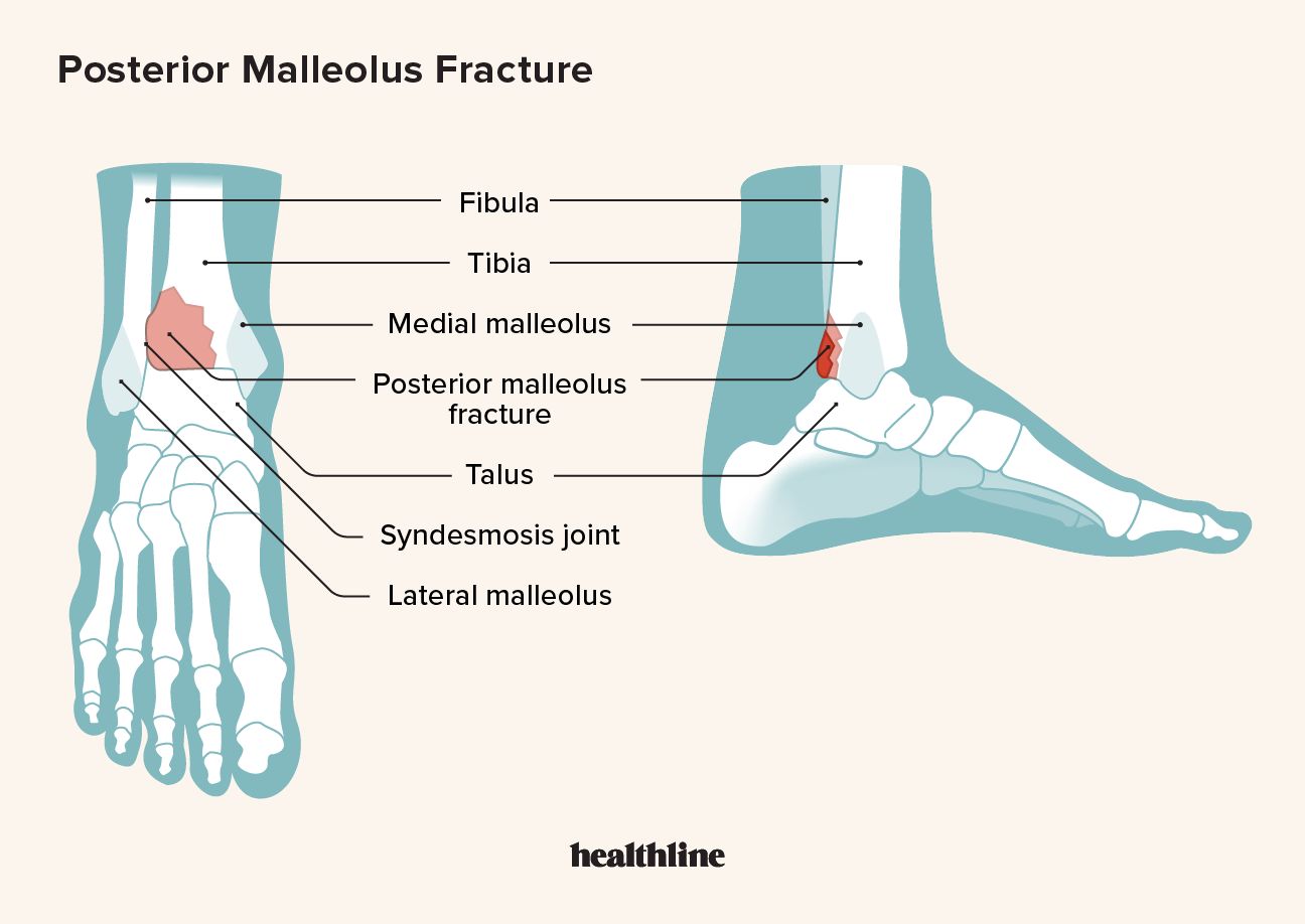
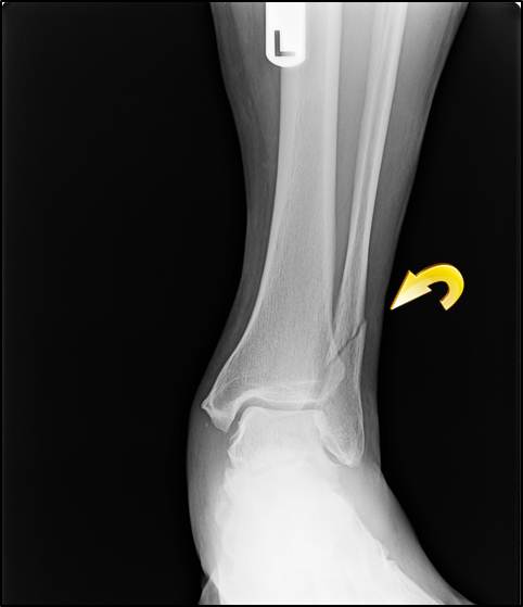
Left Ankle Fracture and Internal Fixation
4.7 (791) · $ 21.99 · In stock

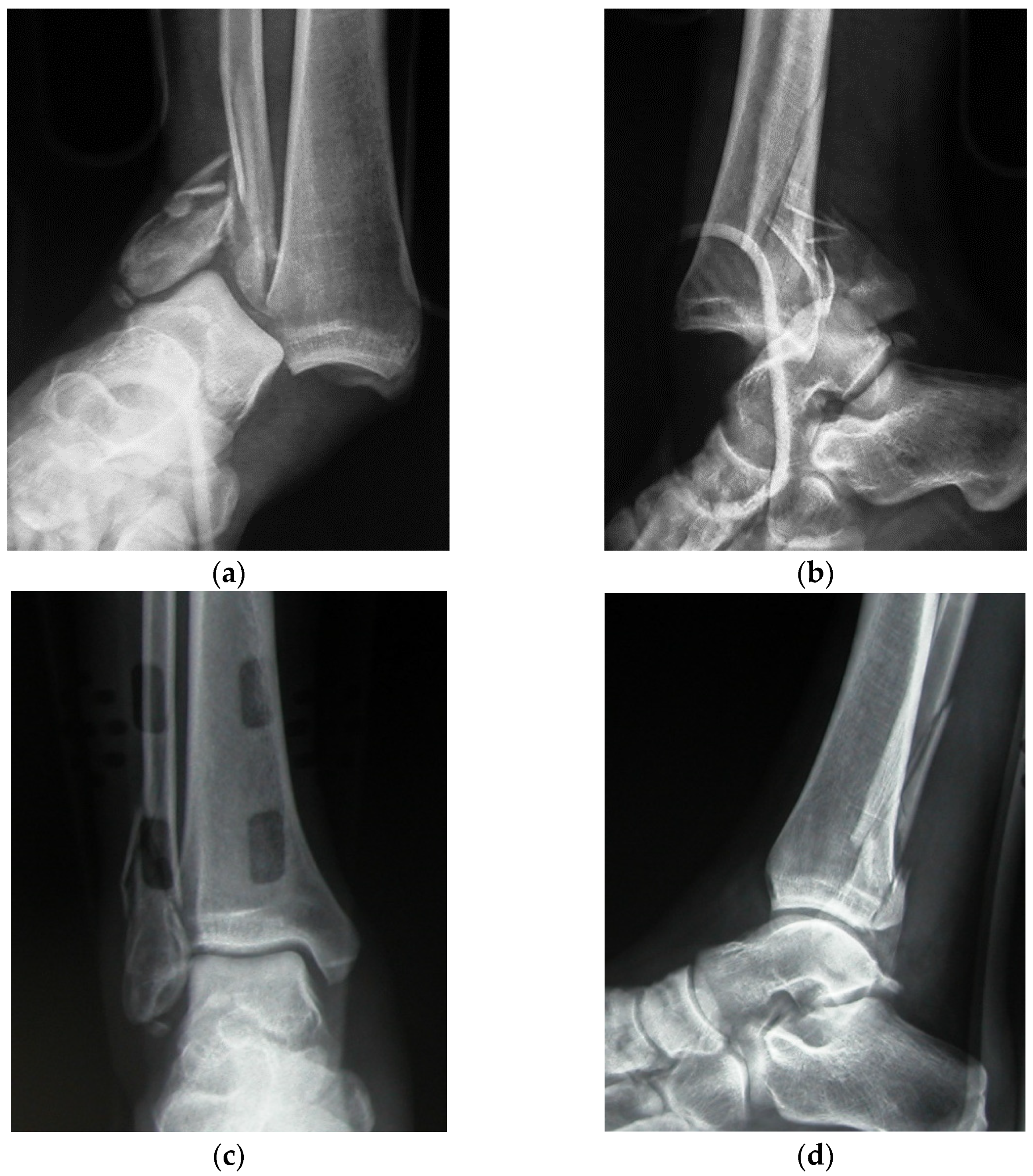
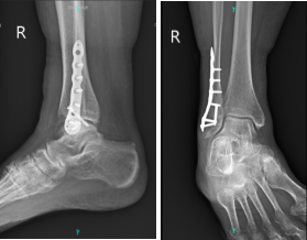
This exhibit features three radiological colorizations showing an ankle fracture and subsequent internal fixations. The first image depicts a fracture of the distal fibula, fracture of the distal tibia, and disruption of the ankle mortise. The second shows reduction of the fracture fragments with the placement of a fibular plate and multiple screws. Lastly, the third image illustrates fusion of the tibiofibular joint with a syndesmotic screw to reduce widening of the ankle mortise.
JFMK, Free Full-Text

x-ray colorization Archives - Anatomical Justice

Ankle arthrodiastasis in conjunction with treatment for acute
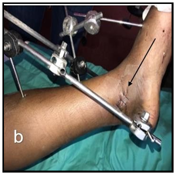
Technique and outcome of ankle fractures treated with Ilizarov

Medical Illustration Open Reduction Internal Fixation of Right
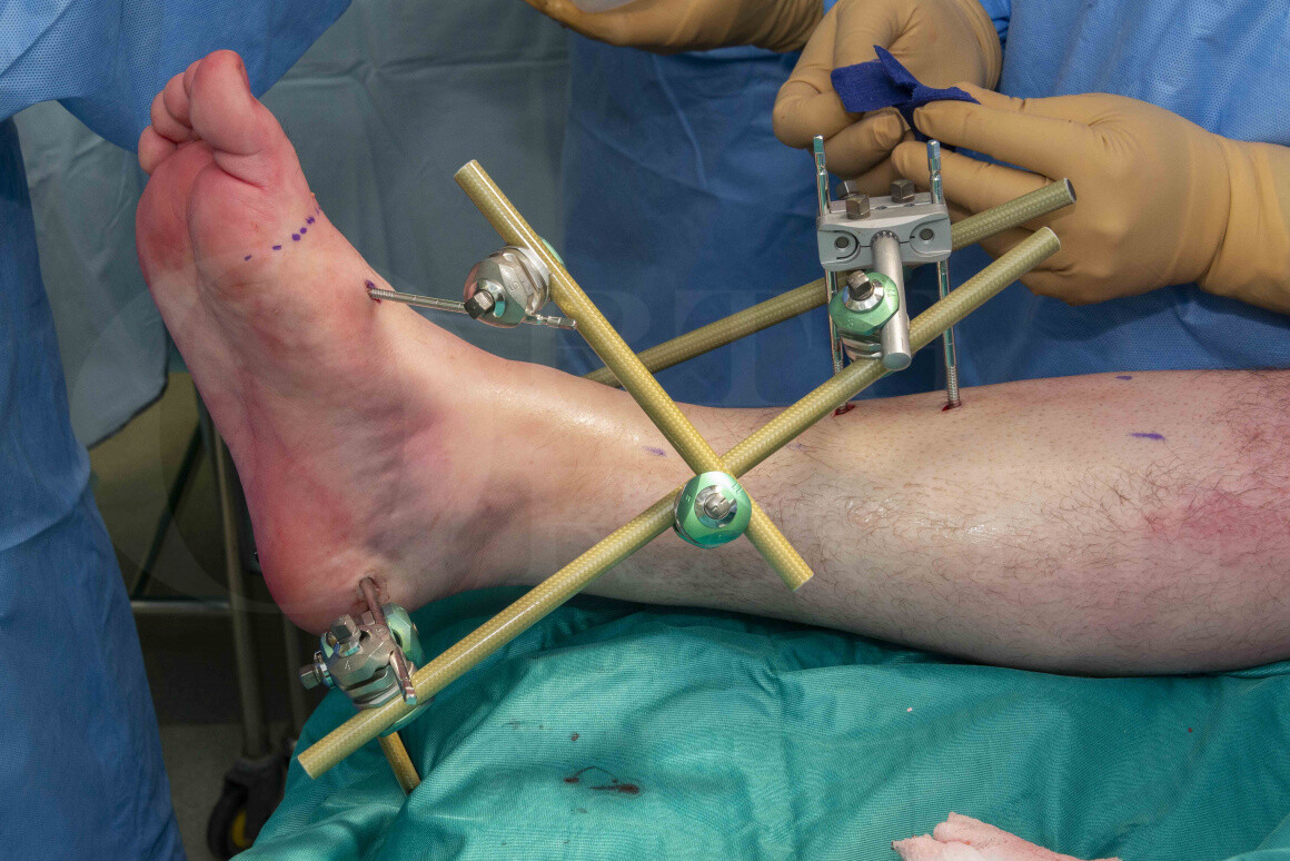
Ankle fracture: Stabilisation with Hoffmann 3 Ankle-spanning
Case Study: Open Reduction and Internal Fixation of Distal Fibula

Maisonneuve fracture - Wikipedia
:max_bytes(150000):strip_icc()/Ankle-Fracture-NYC-Medial-Malleolus-Avulsion-1-56a315cf3df78cf7727bbaa2.jpg)
5 Kinds of Medial Malleolus Ankle Fractures

Early Weight-Bearing Following Ankle Fracture ORIF
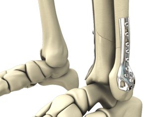
Intraoperative views of fractured left fibula and medial malleolar fracture fixated with plate and screws
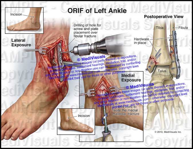
ORIF of Left Ankle

Ankle Joint Art Print By Sebastian Kaulitzki/science Photo, 45% OFF
Open Reduction and Internal Fixation of the Ankle Kentucky
What Ankle Fracture Treatment is Right for You?

Arthrex - Internal Fixation of Ankle Fractures