10.1055/b-0034-74149 Male Foot Fig. 17.1 Neonate Fig. 17.2 Neonate Fig. 17.3 3-month-old Fig. 17.4 3-month-old Fig. 17.5 6-month-old Fig. 17.6 6-month-old Fig. 17.7 9-month-old Fig. 17.8 9-month-ol…

Male Ankle Radiology Key
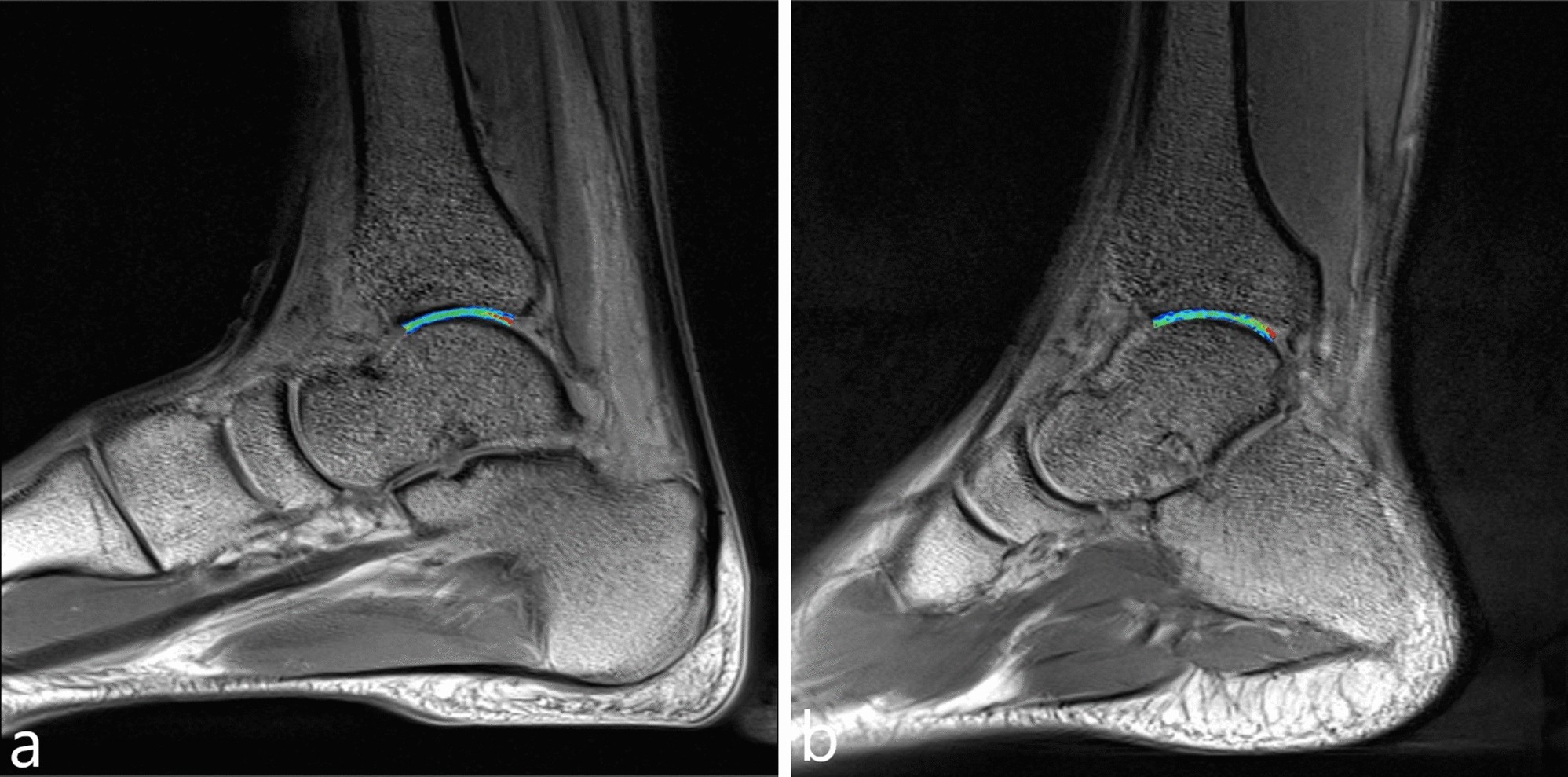
T2 mapping for quantitative assessment of ankle cartilage of weightlifters

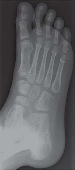
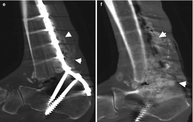
Radiographic abnormalities of foot and ankle. a. Antero-posterior and

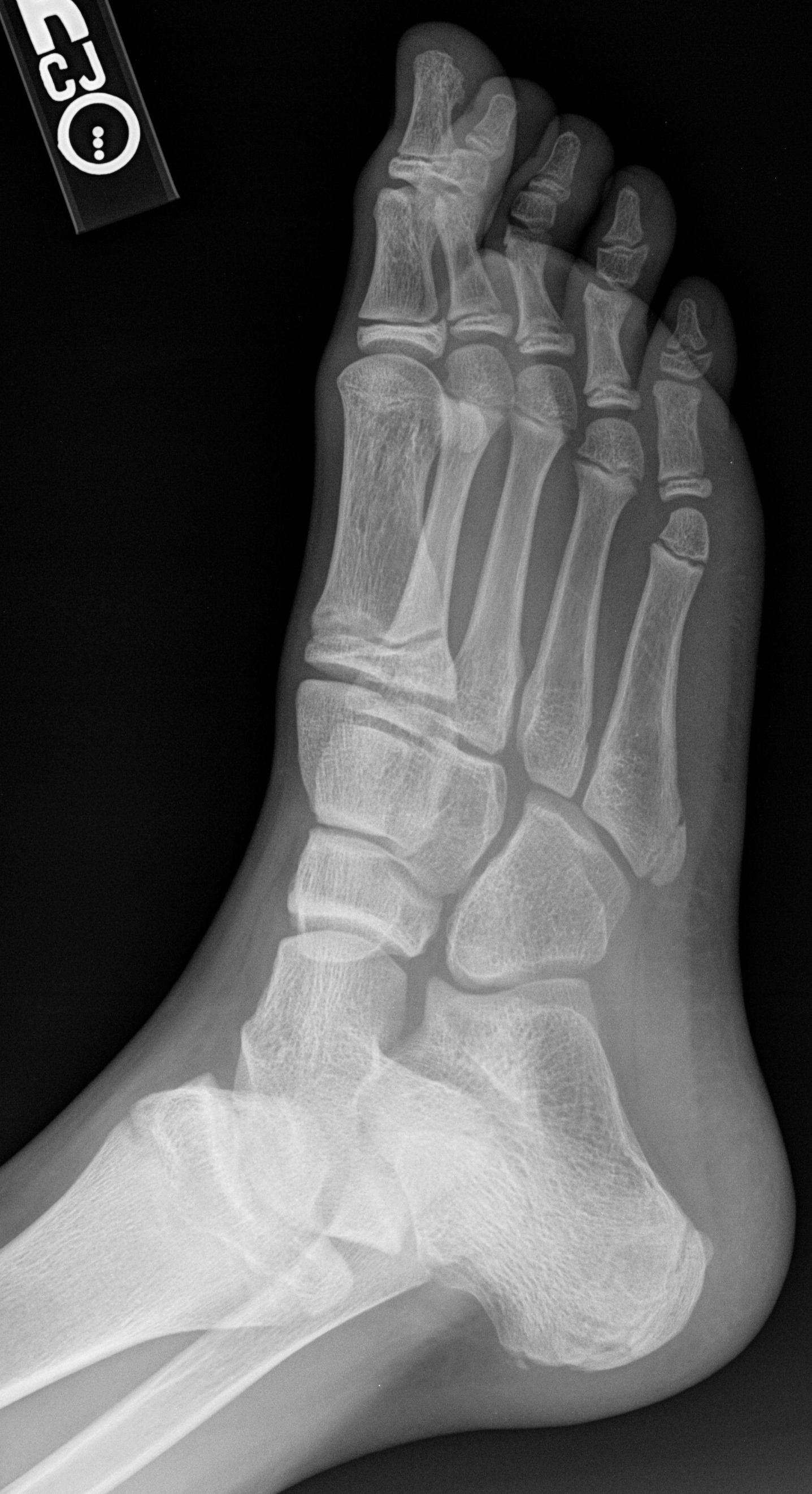
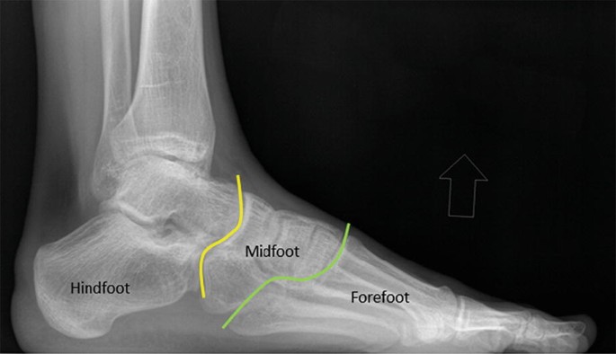
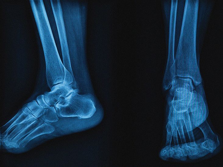
Anatomy of Foot X-rays
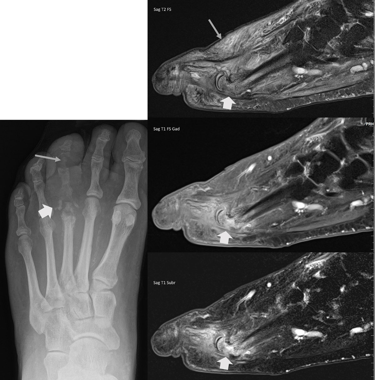
Imaging of the Diabetic Foot - Journal of the Belgian Society of Radiology

Pes planus, Radiology Reference Article
Radiological anatomy: X-ray, CT, MRI
Imaging of Soft Tissue Injuries of the Foot and Ankle

Part 2: Bone Tumors
The Diabetic Foot

Pes cavus, Radiology Reference Article

Ankle x-rays - Don't Forget the Bubbles
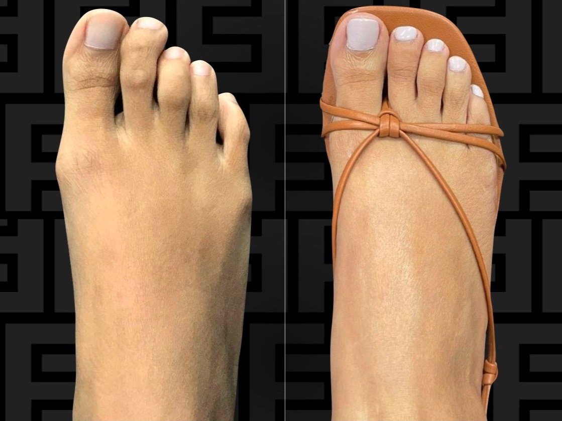
Foot anatomy: Pictures, models, and common conditions of the foot