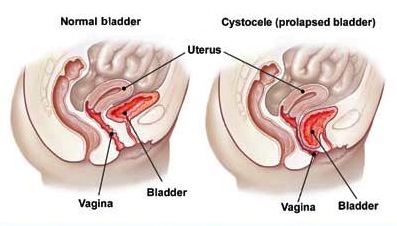
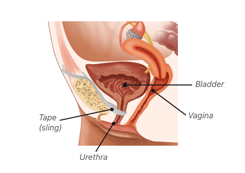
Prolapsed Bladder (Cystocele) Surgical Repair with a Vaginal Slin
4.8 (76) · $ 7.50 · In stock

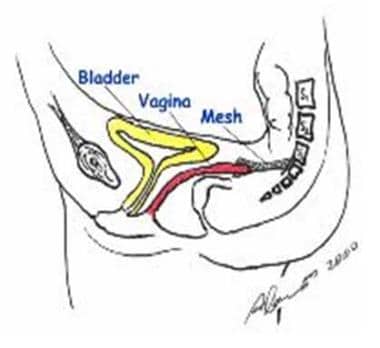
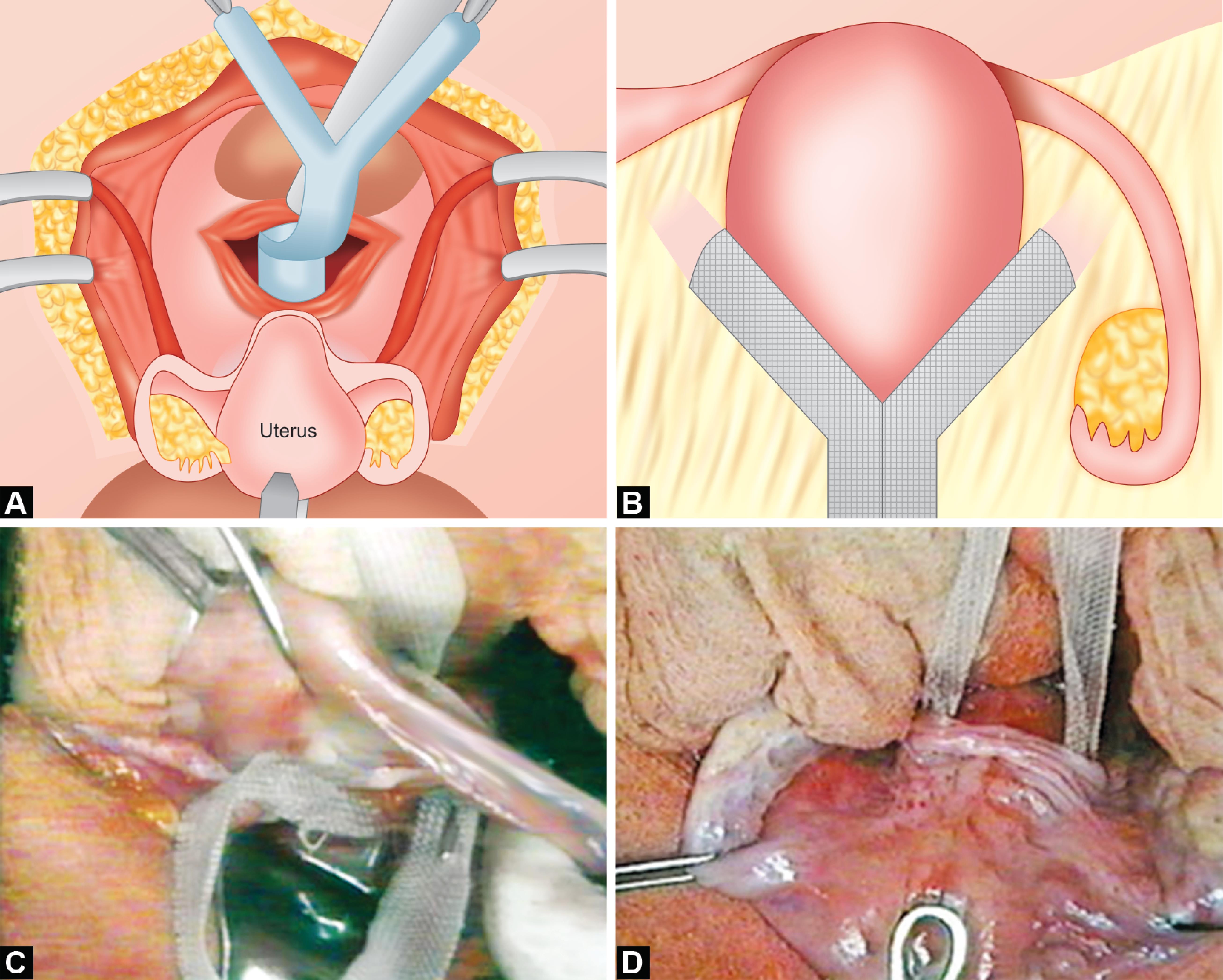
Animation describing normal female pelvic anatomy compared to a patient with a prolapsed bladder and the repair of the prolapsed bladder with a vaginal sling. Incisions made on either side of groin. Sling is inserted with Monarc needle. The mesh is hooked to the needle and pulled back out of the groin incision. Cystoscope placed within the urethra to confirm no bladder injury. Ends are cut, plastic is pulled off and incisions are glued. Normal flow with sling in place.
Pelvic Organ Prolapse Treatment & Management: Medical Therapy
Dr. Lawrence Lin, MD, FACOG
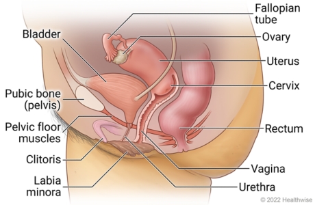
Pelvic Organ Prolapse
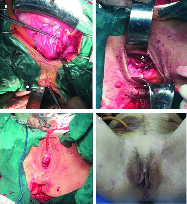
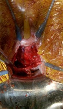
Vaginal Hysterectomy, Uterosacral Ligament Suspension, Anterior
Prolapse Surgery - RYC®
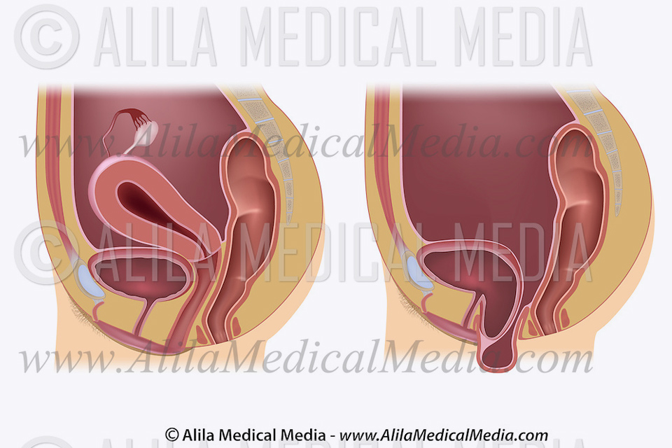
Pelvic Organ Prolapse
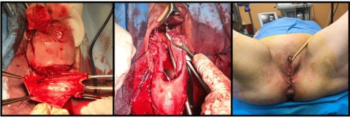
Pelvic Organ Prolapse, Riachi Surgery
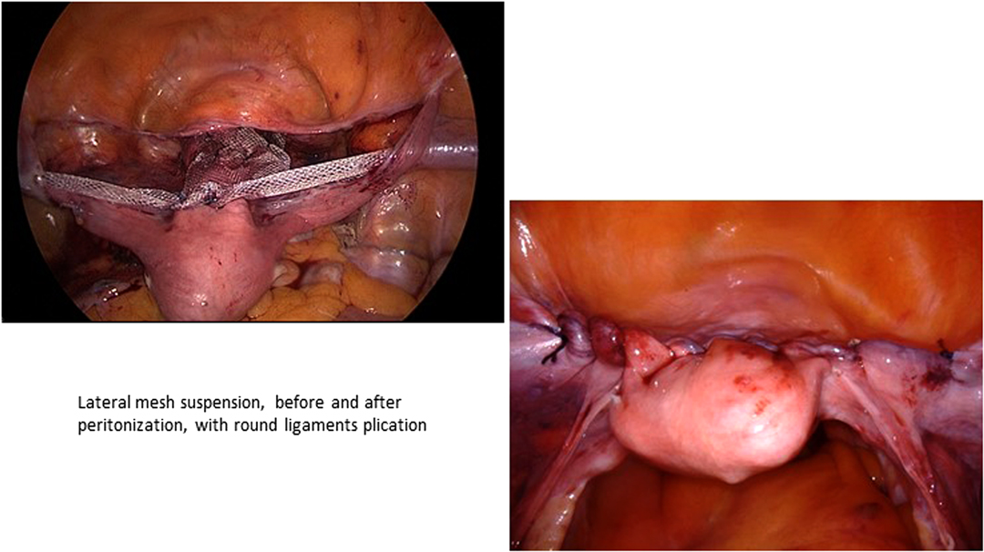
Frontiers Laparoscopic Lateral Suspension (LLS) for the
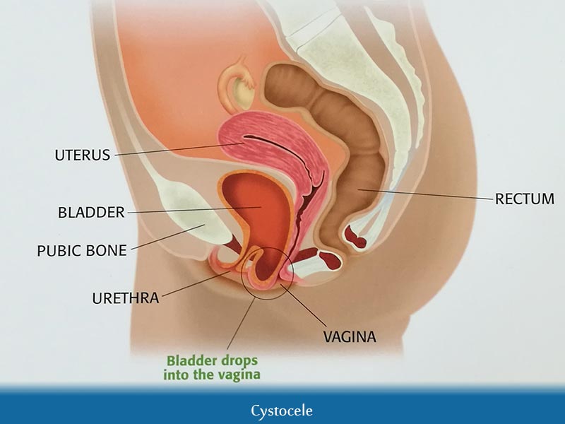
Cystocele Repair: Overview, Technique, Periprocedural Care
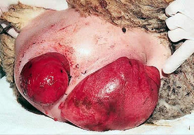
Stasis ulcer and hydronephrosis after severe genital prolapse: a
Pelvic Organ Prolapse: From Basics to Newer Evolutions in Surgical

Pelvic Organ Prolapse - Dr. Nitu Bajekal

Pelvic Organ Prolapse
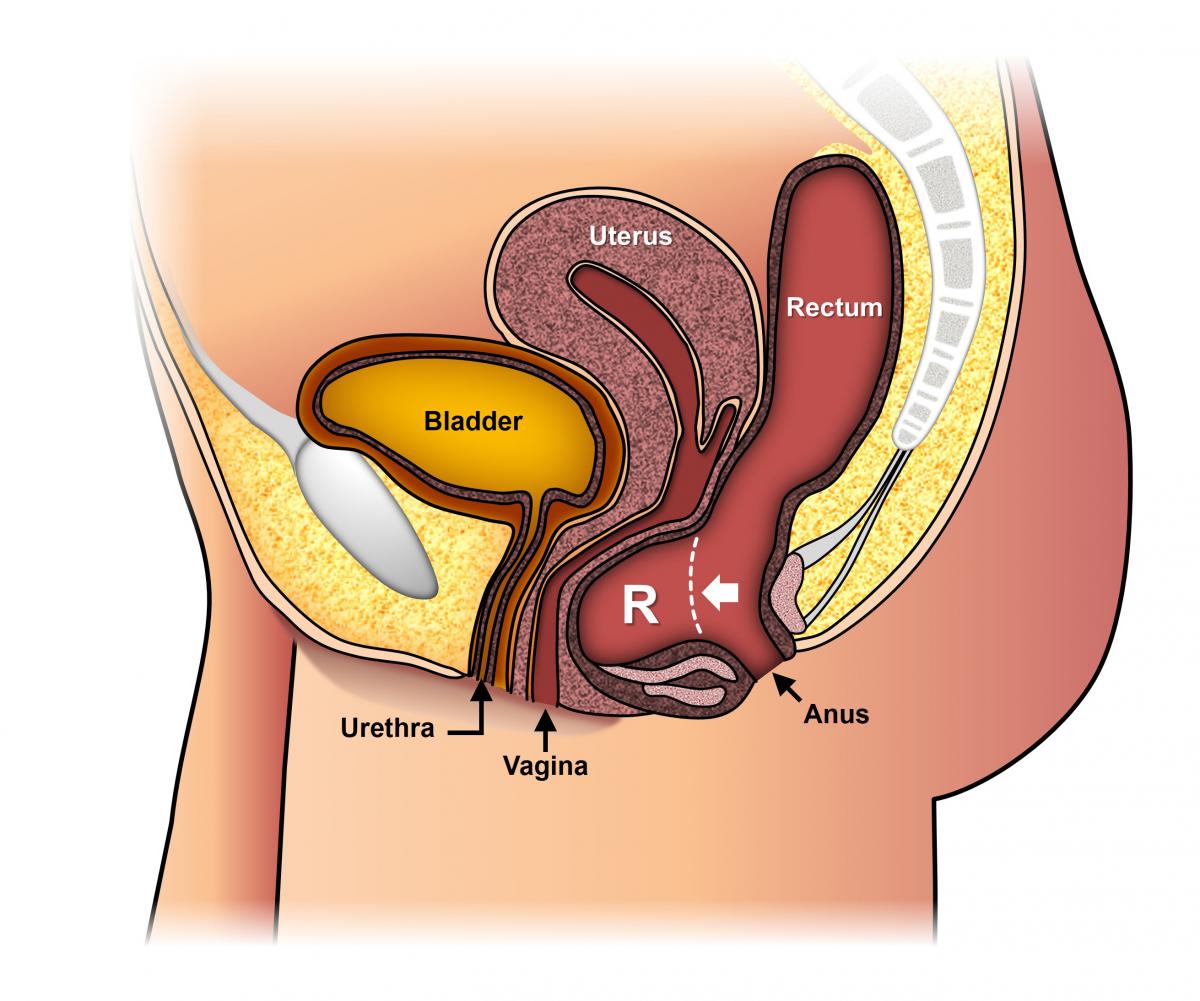
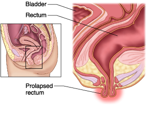
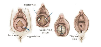
Rectocele