Histology, microscopy, anatomy and disease: Week 3: 2.1
4.8 (491) · $ 11.50 · In stock


Histology, microscopy, anatomy and disease: Week 3: Figure 12 a) The human nervous system showing the CNS and PNS. (b) A diagram of a small peripheral nerve showing the mixture of nerve
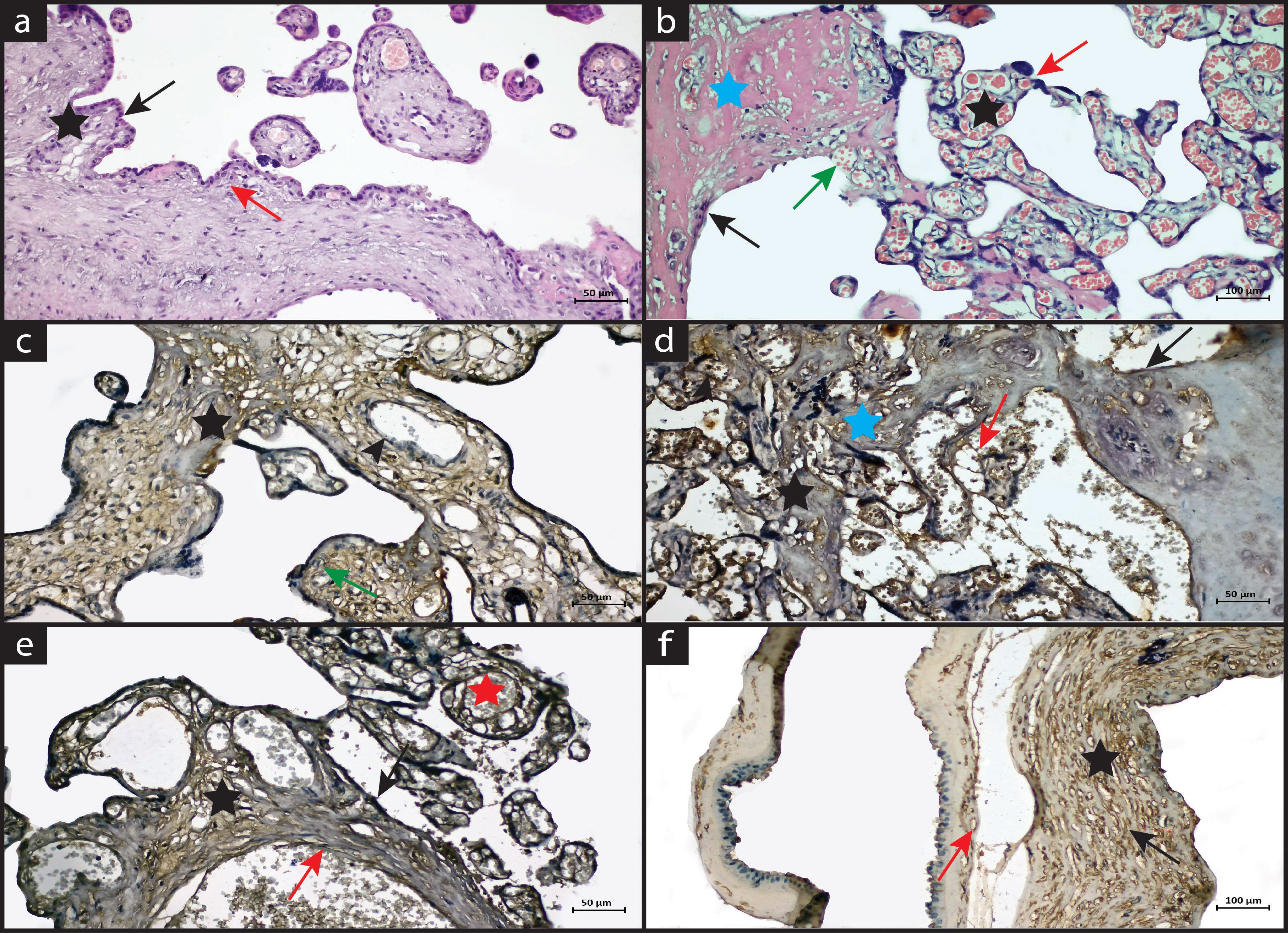
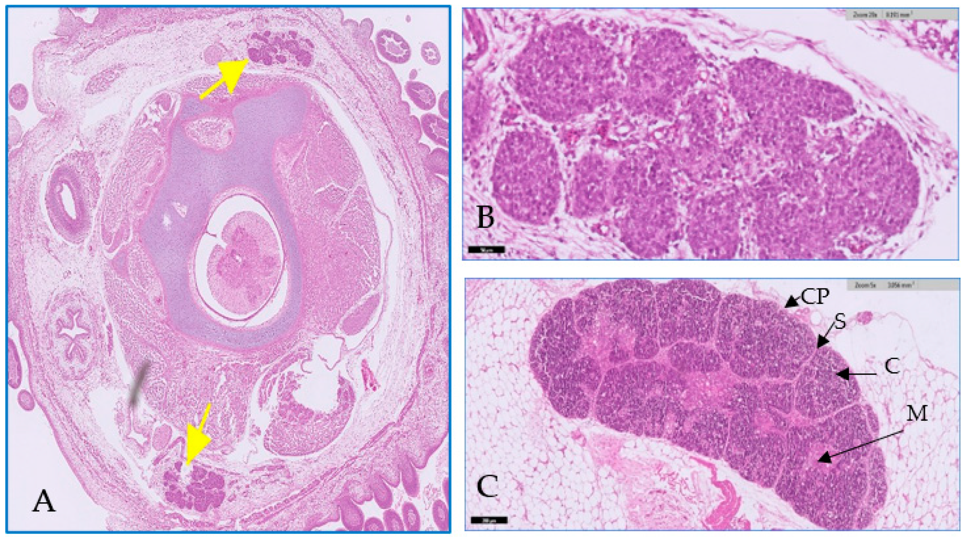
The Investigation of Caspase-3 and Tumor Necrosis Factor-Alpha Expression in Placentas of Patients with Preterm Premature Rupture of Membranes
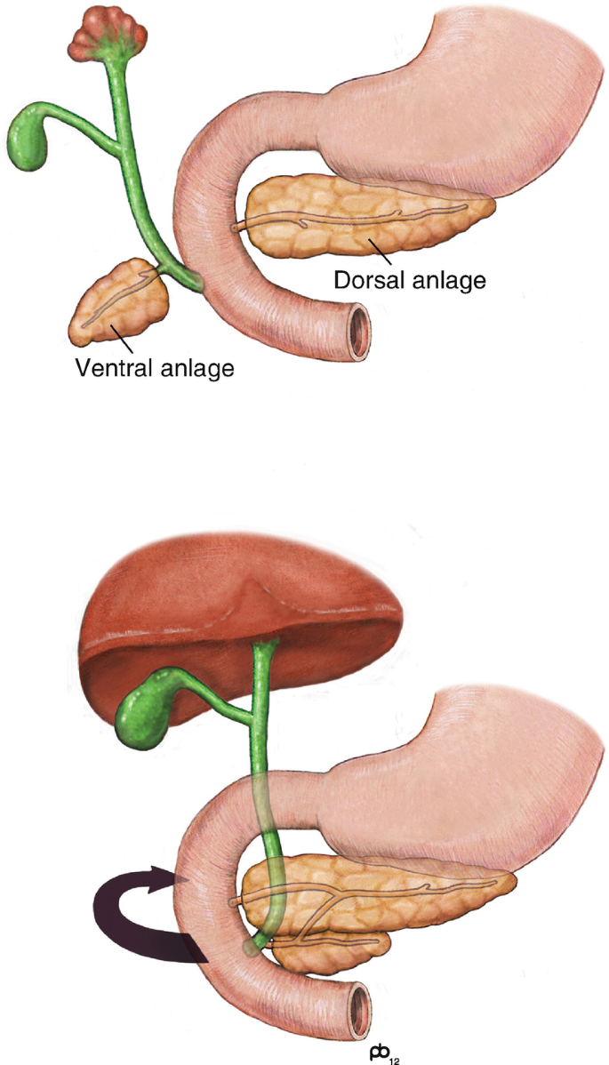
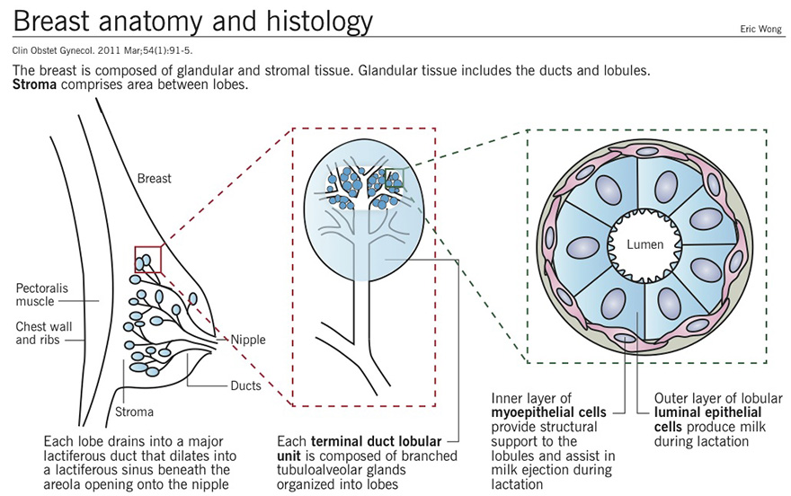
Embryology, Anatomy, and Histology

Infection of 3D Brain Organoids with Human Pathogenic Viruses Under Biosafety Level-3 Conditions with Subsequent Inactivation to Study Viral Replication, Pathomechanisms, and Other Viral Infection-Mediated Effects
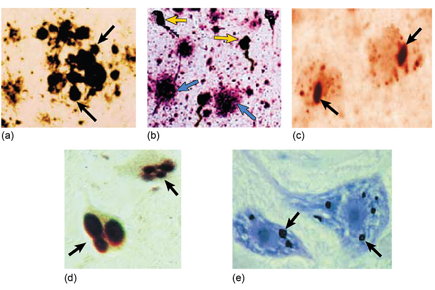
Histology, microscopy, anatomy and disease: Week 4: Figure 7 Protein aggregates in brain cells associated with neurodegenerative disease. Arrows highlight (a) extracellular plaques in prion disease, (b) extracellular plaques (blue) and neurofibrillary
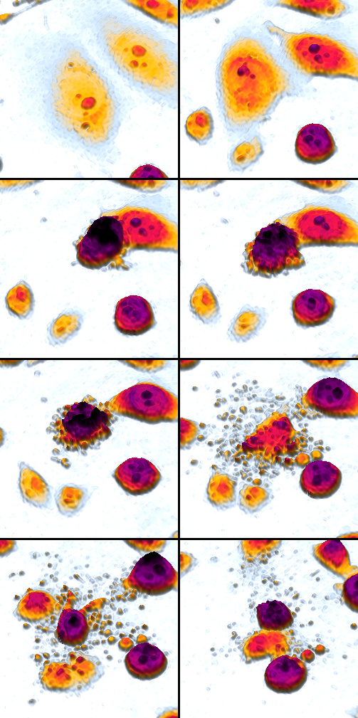
Apoptosis - Wikipedia
Toxics, Free Full-Text

Granuloma - Wikipedia

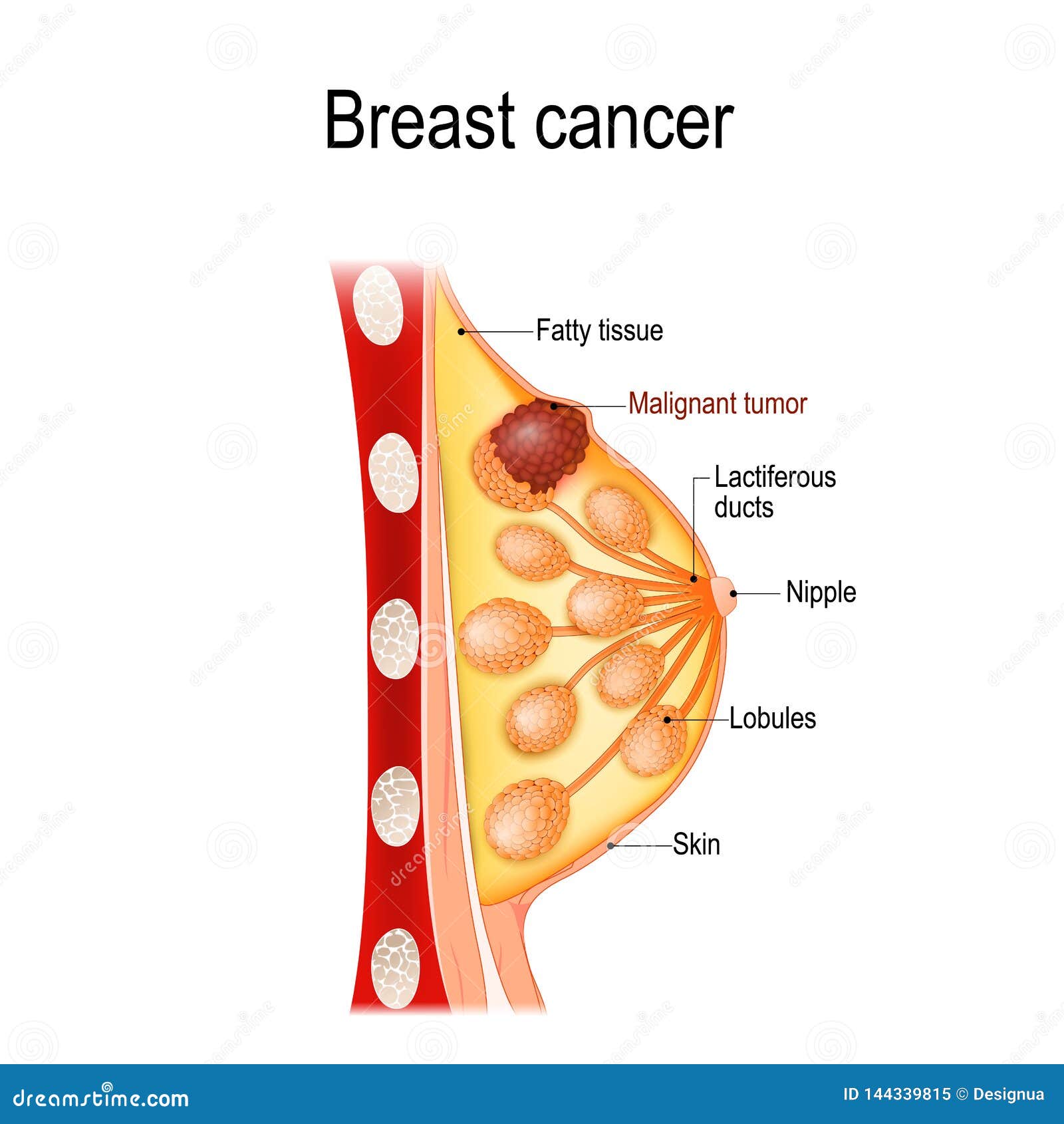
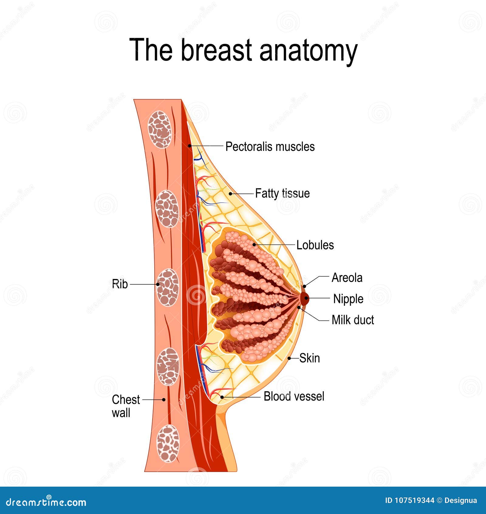
High quality, high discounts Breast Anatomy: Milk Ducts, Tissue

High-speed light-sheet microscopy for the in-situ acquisition of volumetric histological images of living tissue
CAP 2013 Surveys and Anatomic Pathology Education Programs

Chagas disease - Wikipedia
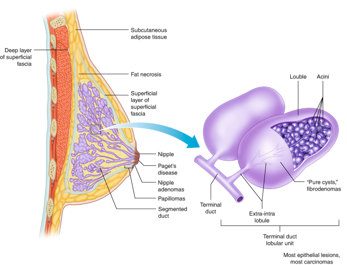
High quality, high discounts Breast Anatomy: Milk Ducts, Tissue