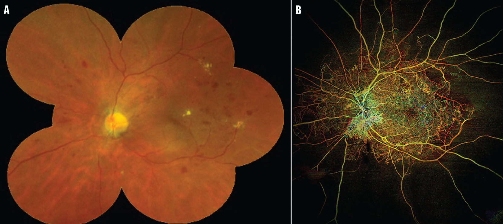
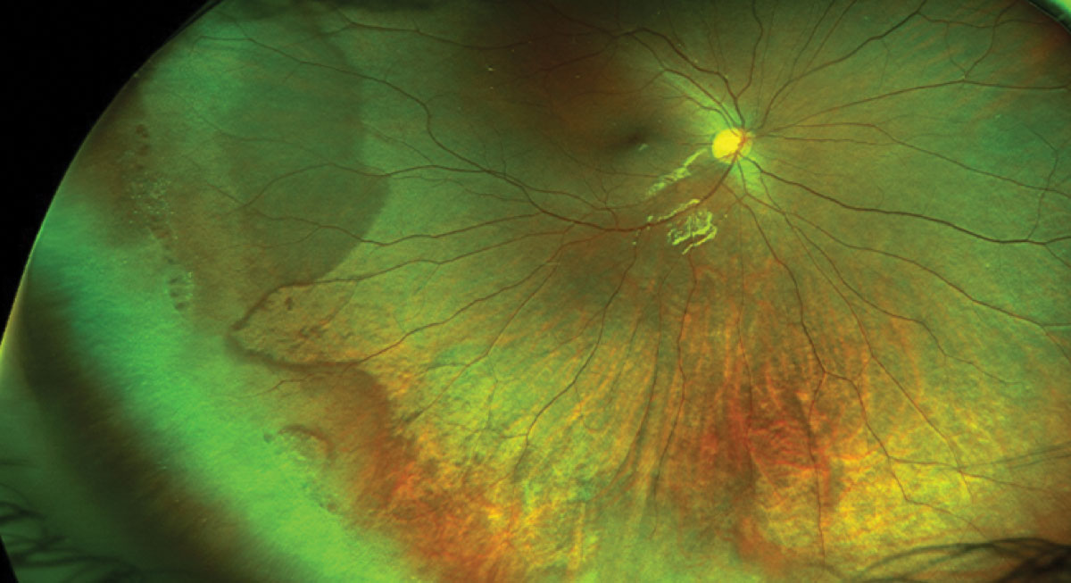
Ultra-wide-field fundus photographs and ultra-wide-field
4.5 (341) · $ 24.99 · In stock

Download scientific diagram | Ultra-wide-field fundus photographs and ultra-wide-field fluorescein angiographic imaging of ocular toxocariasis. (A) A granuloma with mild vitreous opacity. (B) A tractional retinal fold with localized tractional retinal detachment. (C) Diffuse peripheral vascular leakage. (D) A prominent optic disc leakage. from publication: The Clinical Characteristics of Ocular Toxocariasis in Jeju Island Using Ultra-wide-field Fundus Photography | Toxocariasis, Ocular and Photography | ResearchGate, the professional network for scientists.

Ultra-wide field color fundus photograph of the right (A) and left (B)
Widefield OCTA: A New Way to Stage Diabetic Retinopathy - Retina Today

PDF) The Clinical Characteristics of Ocular Toxocariasis in Jeju Island Using Ultra-wide-field Fundus Photography
Ultra-widefield Imaging Ideal for Monitoring Myopic Maculopathy
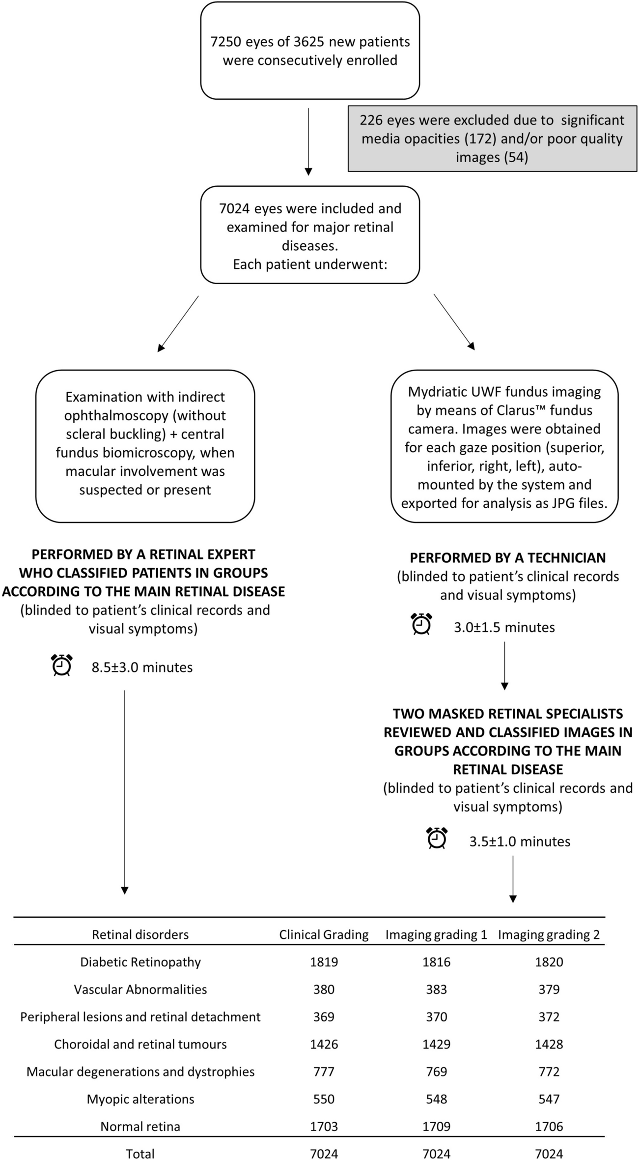
Ultra-wide-field fundus photography compared to ophthalmoscopy in diagnosing and classifying major retinal diseases

Sang-Yoon Lee's research works Gachon University, Seongnam-si (kyungwon) and other places

How these Australian ophthalmologists maximise Optos ultra-widefield retinal imaging - Insight
How ultra-widefield imaging is changing our view of DR
Wide-field Imaging of Retinal Diseases - touchOPHTHALMOLOGY